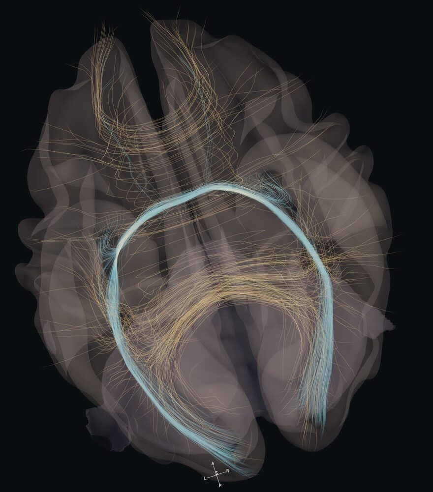
Commissural bundles on the reference brain, viewed from above.
Hodos

The brain, as pathways. Explained.

Cortex, white matter and brain networks in one 3D atlas, taught as relationships you explain, not pictures you recognise.

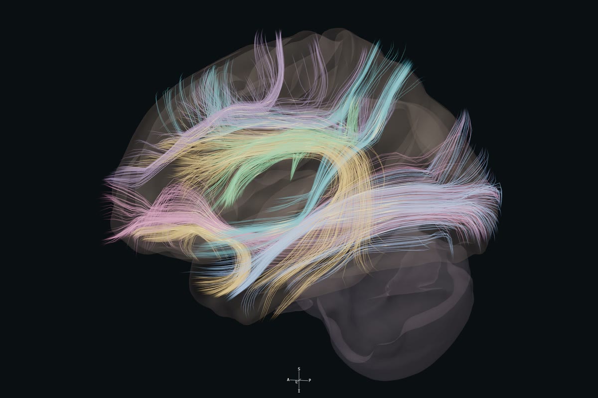
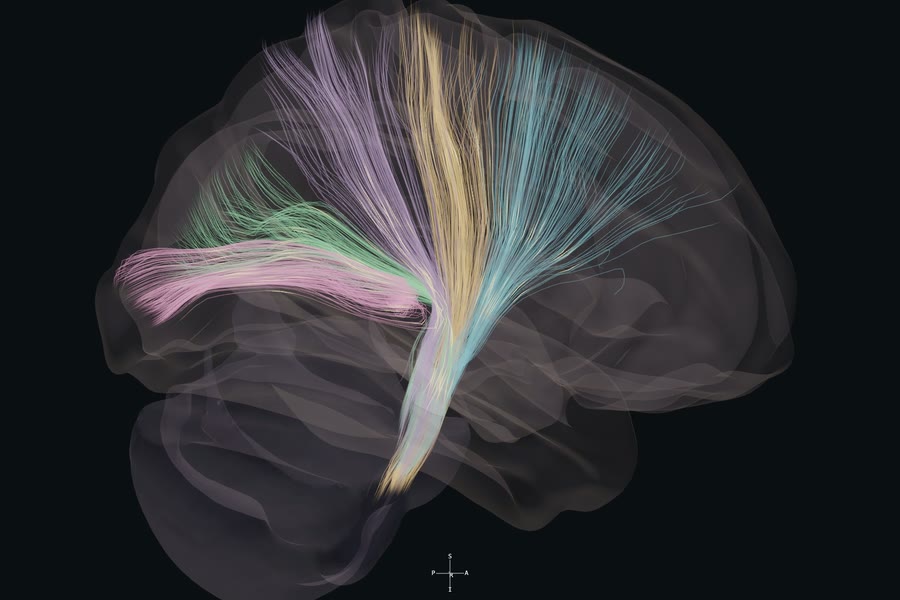
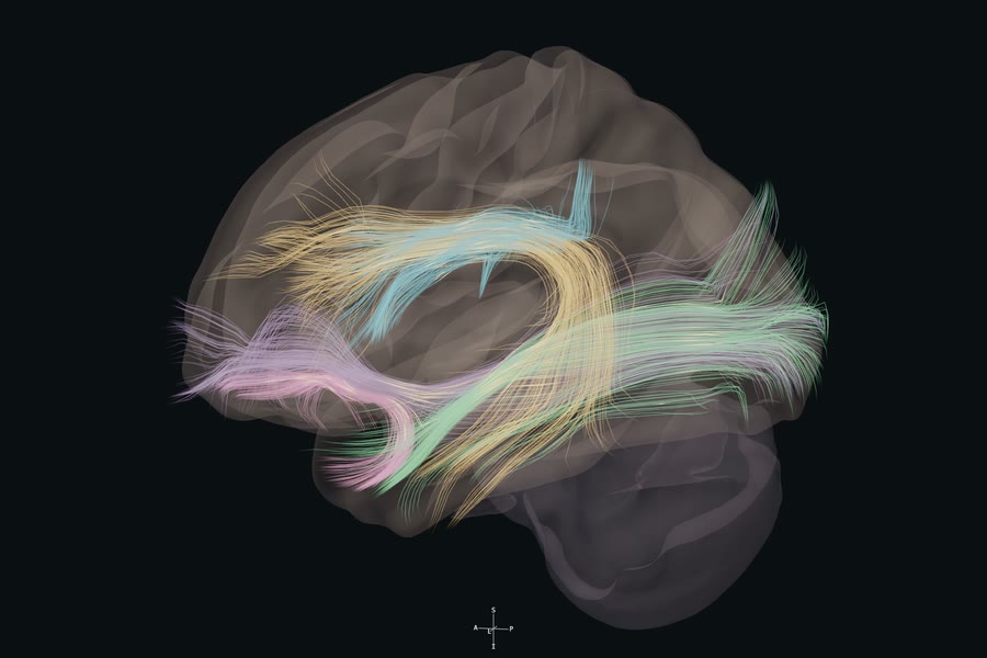
Commissural bundles, superior view, captured from the live atlas. The loop continues through association and projection pathways.

14
lessons for neurosurgical residents
85
relationships, each with a question and its sources
4
published reference datasets, credited
Free
in the browser, no registration. Educational reference, not navigation.

Four families, marked by the way their fibres run.

Red left to right

Green front to back

Blue up and down

The tractography direction code. Here it marks a family’s dominant direction; the atlas renders keep their own pigments.

Fourteen lessons. Orient, compare, explain.

Each lesson runs on the live atlas. You locate the structures, put the neighbours in the same view, and answer a resident-level question before the explanation and its sources are revealed. Lessons run 8 to 22 minutes, depending on how many relationships each one covers.

All fourteen lessons, with their questions and sources

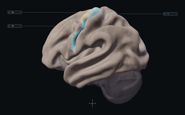
The central region lesson in the atlas: areas 4, 3b and 6d lit on the left hemisphere, the Orient step with its three targets, and the lesson question beside the anatomy.
A lesson as it runs, recorded from the live atlas: select the structures, turn the brain, then move from Orient to Compare to Explain. The ring marks the pointer.

The other eleven

  1. Read the evidence

    A highlighted parcel, a nearby tract and weakness appear in one report. What does each actually establish?

    8 min
  2. When the tractogram is incomplete

    A tract looks short and sparse. How do you describe the discrepancy without declaring it absent?

    8 min
  3. Optic radiation & temporal stem

    An expected anterior loop appears short in a reconstruction. What would you investigate first?

    12 min
  4. Insula & central core

    A left insular lesion extends toward the lentiform complex despite preserved naming. Which observations are still needed?

    12 min
  5. Attention & spatial awareness

    A person omits items on the left during search despite useful hand strength. How would you localize the question?

    18 min
  6. Language across pathways

    Speech is fluent but meaning-related substitutions occur, with repetition better preserved than comprehension. Which relationships matter?

    22 min
  7. Salience, insula & control

    After a frontoinsular procedure, redirecting behaviour toward relevant events is difficult. What must a useful explanation include?

    18 min
  8. Internal capsule & thalamic radiations

    A left deep lesion is followed by right face, arm and leg weakness with hemisensory loss but no visual-field symptom. Which capsular part leads?

    16 min
  9. Corpus callosum & cingulum

    Which callosal segment and neighbouring systems matter when a posterior disconnection hypothesis affects visual transfer but spares basic motor output?

    16 min
  10. Ventral stream & the temporal stem

    A resident must explain a semantic or phonological deficit near the anterior temporal lobe. Which pathway relationship should be examined first?

    14 min
  11. Brainstem & cranial nerve corridors

    Ipsilateral facial weakness accompanies contralateral body weakness. Which cranial nerve level and long tract relationship come first?

    16 min

Then practise the reasoning.

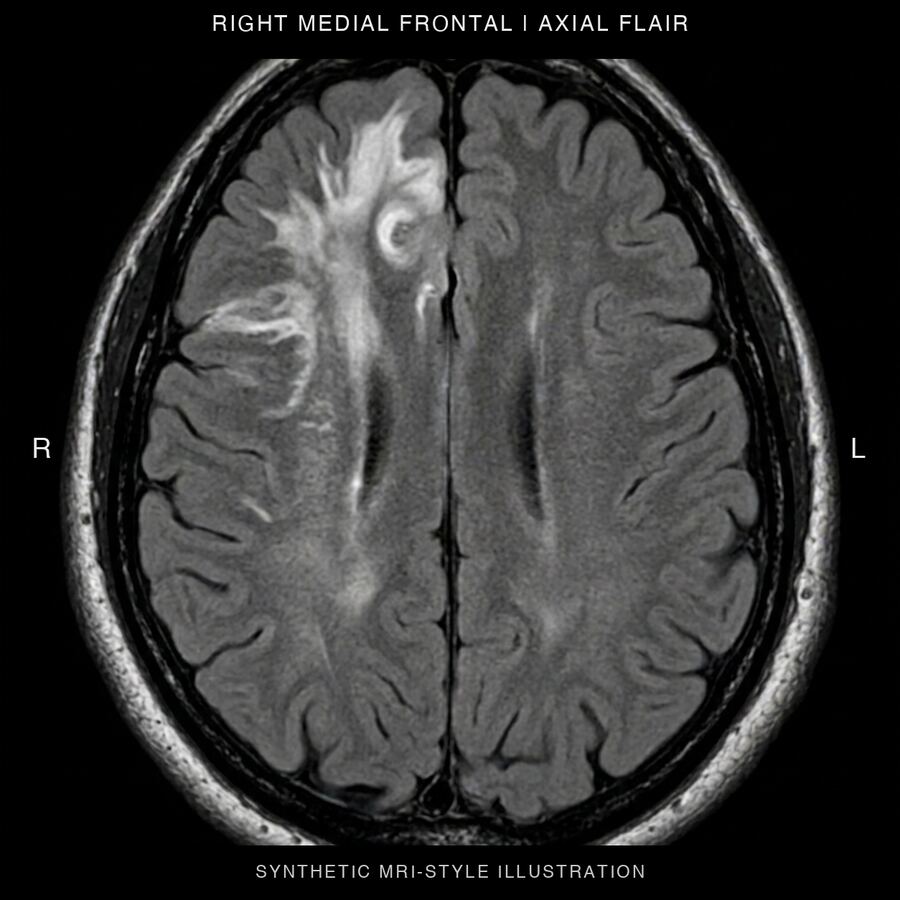
Synthetic MRI-style plate for the fictional right medial frontal Case Conference vignette.

Case Conference

Four fictional glioma teaching cases for senior residents. Read the vignette, commit to an anatomical account, then compare it with the debrief. Synthetic MRI-style plates, no patient data.

Open the Case Conference

Pilot. Debriefs are awaiting independent clinical review.
MIPS exercise

A fictional deep frontal target and two illustrative access corridors, worked through on the reference atlas. Orient the anatomy, compare the corridors, then explain what changes between them.

Try the MIPS exercise
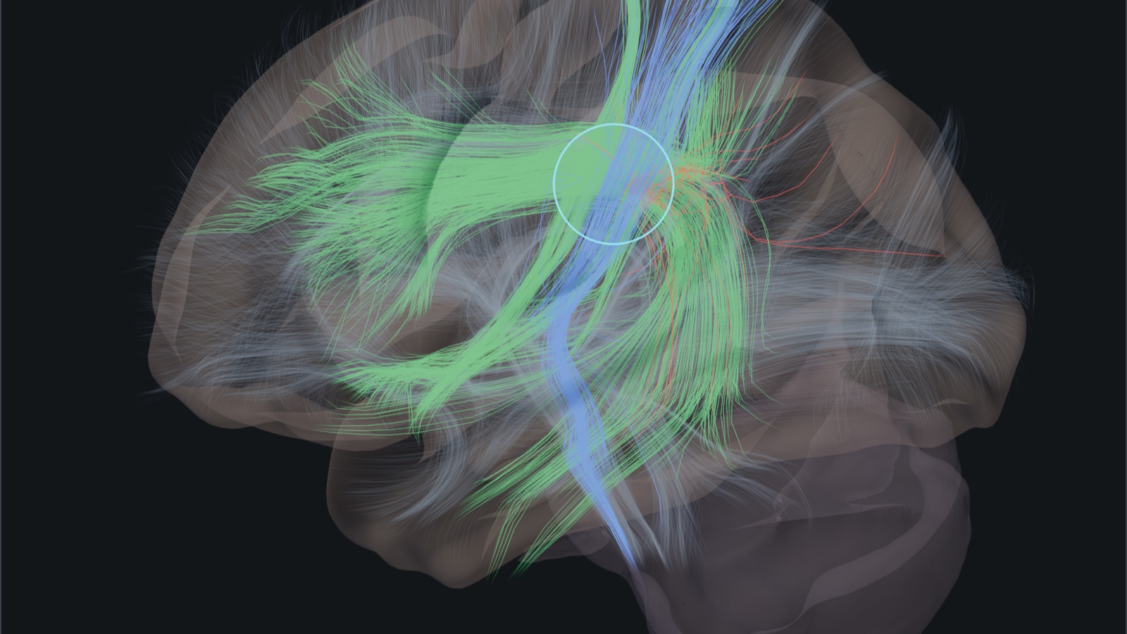
The lesion lab: a translucent sphere in the white matter of the left hemisphere, with the sampled streamlines it cuts drawn along their whole length on the group reference brain.

Lesion lab

Drag a lesion sphere through group-average white matter and see which sampled bundles it cuts. For each bundle, the lab quotes what the author’s teaching notes say injury to it causes, with the sources, or says no quoted evidence.

Open the lesion lab

Group reference, not this patient. Educational draft awaiting independent clinical review.

Made by a neurosurgeon who teaches residents.

Hodos was built by Dr. Erion de Andrade, neurosurgeon in Porto Alegre with skull base and neuro-oncology fellowships at Cleveland Clinic and Emory University, and residency preceptor at Hospital São José, Santa Casa de Porto Alegre. Hodos is the Greek word for path, the root of hodotopy: function lives in the pathways as much as in the cortex.

Published data, cited on every lesson.

  • HCP S1200 group surface
  • HCP-MMP1 cortical parcels
  • HCP1065 sampled tractography
  • Melbourne subcortex atlas

Licences and limits are on the sources page.

Start with one relationship.

About 20 minutes on the live atlas. Orient the central region, compare its banks, explain the deficit.

Start a lesson
Left lateral atlas view with area 4 highlighted in ice and areas 3b and 6d in sand, with labelled leaders and orientation axes.
Left lateral view. HCP-MMP1 parcels on the HCP group surface.

Two banks of the central sulcus

Compare area 4 with area 3b across the central region. Area 6d adds premotor context in front.

Look for

Which bank lies anterior to the sulcus, and which lies posterior?

These group parcels orient the relationship. Their outlines do not establish an individual's functional boundary.

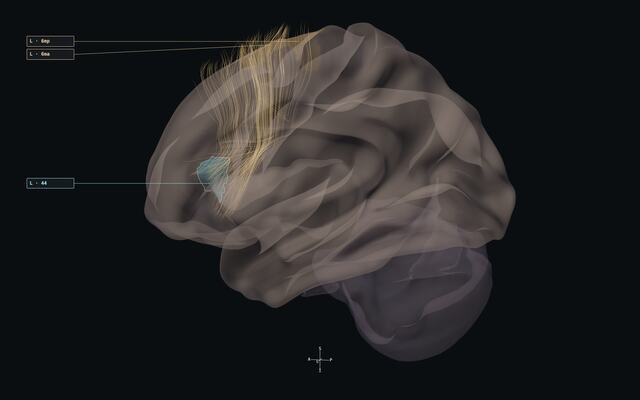
Left frontal aslant tract sample seen through translucent cortex, with atlas parcels 44, 6ma and 6mp labelled.
Left lateral view. HCP1065 sampled pathways with HCP-MMP1 reference parcels.

From lateral to medial frontal cortex

Follow the oblique frontal aslant sample between inferior frontal and medial superior frontal references.

Look for

Find parcel 44, then compare it with medial premotor references 6ma and 6mp.

Sampled tractography illustrates a course. It does not certify individual fibre terminations or language function.

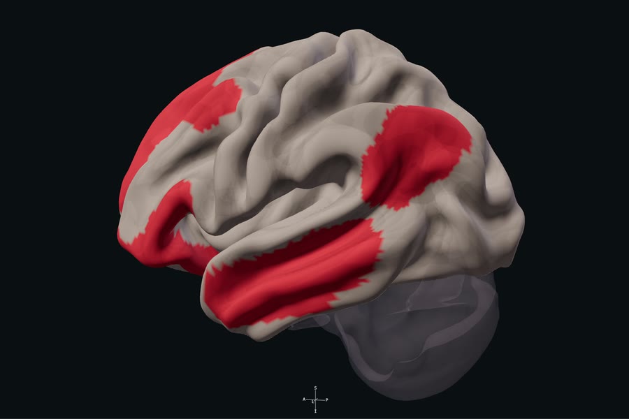
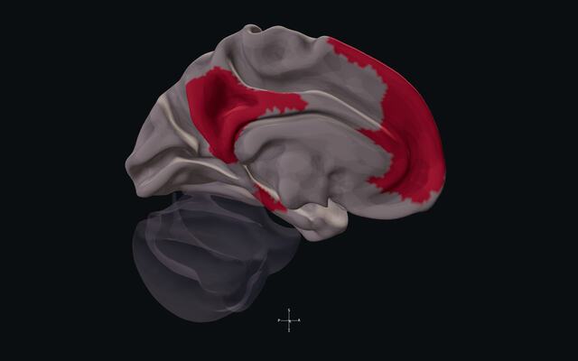
Left medial atlas surface with separated default mode network territories shown in the red Yeo-7 group label.
Left medial view. Yeo-7 default mode network on the HCP group surface.

A network across separated territories

Trace the anterior and posterior medial territories, then rotate the atlas to find the lateral parietal component.

Look for

What part of this distributed network would a single lateral view leave out?

Red identifies membership in the Yeo-7 group network. It does not show activation or surgical risk.